

How High Blood Sugar Causes Cellular Damage: A Mechanistic Guide
Persistent high blood sugar damages cells by driving oxidative stress, protein glycation, and maladaptive metabolic pathways that impair repair and trigger cell death. This is not simply an osmotic problem. Elevated intracellular glucose hijacks mitochondria, floods the polyol and hexosamine pathways, activates protein kinase C (PKC), and generates advanced glycation end-products (AGEs) that bind RAGE receptors to amplify inflammation. The result is a self-reinforcing cycle of reactive oxygen species (ROS) overproduction, redox imbalance, and cumulative structural injury across virtually every organ system.
Clinically, the thresholds that define meaningful risk are well established:
- Fasting plasma glucose: above 125 mg/dL signals diabetes-range hyperglycemia
- 2-hour postprandial glucose: above 180 mg/dL indicates impaired glucose disposal
- HbA1c: the ~7% target endorsed by the American Diabetes Association reflects average glucose exposure over roughly three months
- Risk framing: damage is a function of cumulative dose × time — how high glucose climbs and how long it stays elevated both matter independently
Even brief, repeated glucose spikes can initiate biochemical injury before HbA1c rises into the diagnostic range. The pathways described throughout this article are the mechanistic reason why early, tight glycemic control remains the most evidence-backed strategy for preventing long-term complications.
Table of Contents
- Does the duration of high glucose change how much damage occurs?
- What molecular pathways does high glucose activate inside cells?
- What actually happens inside individual cells when glucose stays high?
- How does cellular injury translate to organ-level complications?
- Is the cellular damage from high blood sugar reversible?
- How do clinicians measure glycemic exposure and cellular injury?
- What strategies actually protect cells and reduce complication risk?
- What does the evidence say about antioxidants and SOD supplementation?
- Key Takeaways
- The case for treating glucose toxicity as a systems problem
- Revivify supports cellular health at the source of oxidative stress
- Primary sources and further reading
Does the duration of high glucose change how much damage occurs?
Yes, and the distinction between acute and chronic hyperglycemia shapes both the type and severity of cellular injury. Acute hyperglycemia, such as the glucose surges seen in diabetic ketoacidosis (DKA) or hyperosmolar hyperglycemic state (HHS), produces rapid osmotic stress, inflammatory cytokine release, and acute ROS bursts. These events are dangerous and require urgent care, but the cellular injury they cause can be partially reversible if glucose is corrected quickly.
Chronic hyperglycemia tells a different story. Sustained elevation of fasting and postprandial glucose, reflected in a rising HbA1c, produces cumulative biochemical damage through the pathways described below. According to StatPearls, persistent hyperglycemia is directly linked to retinopathy, nephropathy, neuropathy, and cardiovascular disease — complications that develop over months to years of inadequate control.
The dose × time concept is central to understanding risk. Frequency of glucose spikes, the magnitude of those spikes, and the duration of exposure all contribute independently. HbA1c captures average glucose over roughly 90 days and remains the standard clinical proxy for cumulative exposure. Continuous glucose monitoring (CGM) adds granularity by measuring time-in-range, time-above-range, and glycemic variability — metrics that HbA1c alone can miss.
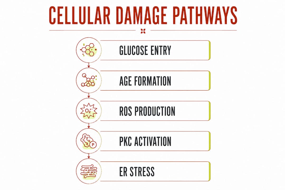
A critical insight from the mechanistic literature: elevated ROS increases insulin resistance, which further raises blood glucose, which generates more ROS. This feedback loop means that even moderate chronic hyperglycemia can accelerate its own damage over time, making early intervention disproportionately valuable.
What molecular pathways does high glucose activate inside cells?
High intracellular glucose activates at least five well-characterized toxic pathways simultaneously. They are not independent; they converge on a shared outcome of redox imbalance, protein dysfunction, and cell death.
AGEs, RAGE, and chronic inflammation
When glucose reacts non-enzymatically with amino groups on proteins, lipids, and nucleic acids, it forms AGEs. These modified molecules accumulate in vessel walls, basement membranes, and connective tissue. When AGEs bind their receptor, RAGE, the result is RAGE-driven inflammatory signaling that activates NF-κB, promotes pro-inflammatory cytokine secretion, and sustains endothelial dysfunction. RAGE activation also amplifies ROS production directly, creating another positive feedback loop between glycation and oxidative stress.
Mitochondrial overload and ROS generation
Under normal glucose concentrations, the mitochondrial electron transport chain (ETC) runs efficiently. Under hyperglycemia, excess glucose metabolites flood the ETC, causing electron leakage and mitochondrial ROS overproduction. This excess ROS damages mitochondrial DNA, oxidizes ETC proteins, and impairs ATP synthesis — a bioenergetic failure that leaves cells unable to meet repair demands. Hyperglycemia is, in this sense, a fundamental disruption of mitochondrial function, not just an excess of substrate.
Polyol pathway, hexosamine pathway, and PKC activation
The polyol pathway becomes significant when intracellular glucose is high enough to saturate normal glycolytic processing. Aldose reductase converts excess glucose to sorbitol, consuming NADPH in the process. Because NADPH is required to regenerate glutathione (the cell’s primary antioxidant buffer), its depletion leaves cells defenseless against ROS. The pathway can metabolize a substantial fraction of intracellular glucose in diabetic tissues, and the resulting NADH overload further stresses the mitochondrial ETC.
The hexosamine biosynthetic pathway diverts glucose toward UDP-GlcNAc, which drives O-GlcNAcylation of transcription factors and signaling proteins. This post-translational modification alters gene expression patterns in ways that promote insulin resistance and inflammation.
PKC activation follows from diacylglycerol (DAG) accumulation, a direct consequence of elevated intracellular glucose. DAG is a physiological PKC activator; in hyperglycemia, chronically elevated DAG drives sustained PKC activity, which phosphorylates the subunits of NADPH oxidase and amplifies ROS production. The DAG-PKC-NADPH oxidase axis has been described as the “dangerous metabolic route in diabetes” in PMC reviews on ROS and cell death.
ER stress and impaired autophagy
The endoplasmic reticulum (ER) handles protein folding and quality control. Chronic hyperglycemia overwhelms ER capacity, triggering the unfolded protein response (UPR). When UPR cannot resolve the protein-folding backlog, it shifts from a protective to a pro-apoptotic signal. Simultaneously, hyperglycemia impairs autophagy, the cellular recycling system that clears damaged organelles and misfolded proteins. Without functional autophagy, damaged mitochondria and glycated proteins accumulate, accelerating cellular senescence and death.
| Pathway | Trigger | Immediate effect | Downstream pathology |
|---|---|---|---|
| AGE/RAGE | Glucose reacts with proteins/lipids | Protein crosslinking, RAGE activation | Inflammation, endothelial dysfunction, fibrosis |
| Mitochondrial ROS | ETC electron leakage under glucose overload | Mitochondrial DNA damage, ATP depletion | Bioenergetic failure, apoptosis |
| Polyol pathway | Aldose reductase activity | NADPH depletion, sorbitol accumulation | Antioxidant deficit, osmotic stress |
| Hexosamine pathway | UDP-GlcNAc accumulation | O-GlcNAcylation of signaling proteins | Insulin resistance, altered gene expression |
| PKC activation | DAG accumulation | NADPH oxidase phosphorylation, ROS burst | Vascular dysfunction, cytokine release |
| ER stress / autophagy impairment | Protein-folding overload | UPR activation, autophagy suppression | Senescence, apoptosis, protein aggregate accumulation |
What actually happens inside individual cells when glucose stays high?
At the cell level, the pathway activity described above translates into three broad categories of damage: loss of redox homeostasis, protein dysfunction, and altered cell fate.
Redox homeostasis fails when ROS generation outpaces antioxidant capacity. Glutathione depletion (via NADPH consumption in the polyol pathway), SOD overwhelm, and PARP activation following ROS-induced DNA strand breaks all contribute. PARP activation depletes NAD+, worsening the redox imbalance and accelerating cell death signaling. This is a well-documented consequence of glucotoxicity at the protein-modification level.
Protein dysfunction arises from multiple post-translational modifications. Glycation produces crosslinked, functionally impaired proteins. O-GlcNAcylation alters transcription factor activity. Succination (from fumarate accumulation) and nitrosylation (from reactive nitrogen species) further modify protein structure and signaling. The cumulative effect is that oxidation disrupts cellular communication at the receptor, enzyme, and transcription-factor level.
Cell fate shifts toward death or senescence. Intrinsic apoptosis proceeds via mitochondrial cytochrome c release and caspase activation. Necroptosis, a regulated form of inflammatory cell death, also occurs in hyperglycemic tissues and amplifies local inflammation. Cellular senescence, where cells stop dividing but remain metabolically active and pro-inflammatory, contributes to tissue dysfunction without immediate cell death.
Certain cell types are particularly vulnerable:
- Retinal pericytes: lack of glucose transport regulation makes them highly susceptible to intracellular glucose overload
- Glomerular podocytes: critical for filtration integrity; podocyte loss is an early marker of diabetic nephropathy
- Schwann cells and peripheral axons: high metabolic demand and limited regenerative capacity make them sensitive to bioenergetic failure
- Vascular endothelial cells: direct exposure to circulating glucose and AGEs; endothelial dysfunction is the earliest vascular lesion
- Pancreatic β-cells: ROS-induced apoptosis in β-cells accelerates insulin deficiency, worsening the hyperglycemic state
How does cellular injury translate to organ-level complications?
The cellular lesions described above map directly onto the clinical complications that define diabetic morbidity. Understanding the cell-to-organ translation helps clinicians anticipate which patients are at highest risk and which screening tests matter most.
Retinopathy begins with pericyte loss from the retinal microvasculature. Without pericyte support, capillary walls weaken, forming microaneurysms. Progressive endothelial dysfunction leads to macular edema and, in proliferative retinopathy, pathological neovascularization. Screening with dilated fundus examination remains the standard of care.
Nephropathy starts with podocyte injury and glomerular basement membrane thickening. Podocyte loss increases glomerular permeability, producing microalbuminuria — the earliest detectable clinical marker. Continued injury progresses to macroalbuminuria and declining GFR. The Cleveland Clinic notes that chronic hyperglycemia damages small blood vessels throughout the kidney, making blood pressure control as important as glucose control in slowing progression.
Neuropathy reflects Schwann cell dysfunction, axonal bioenergetic failure, and impaired nerve repair. Symptoms range from distal symmetric polyneuropathy (burning, numbness, loss of protective sensation) to autonomic neuropathy affecting cardiac rhythm, gastric motility, and bladder function.
Cardiovascular disease is driven by endothelial dysfunction, AGE-mediated arterial stiffening, and oxidative stress-related stroke risk. Macrovascular disease (coronary artery disease, peripheral artery disease, stroke) accounts for the majority of diabetes-related mortality.
Poor wound healing results from impaired neutrophil and macrophage function, reduced angiogenic response, and peripheral neuropathy that delays recognition of injury.
| Organ | Key cell targets | Principal mechanisms | Common clinical markers |
|---|---|---|---|
| Retina | Pericytes, endothelial cells | Microaneurysm formation, neovascularization | Fundus exam findings, visual acuity |
| Kidney | Podocytes, mesangial cells | Basement membrane thickening, filtration loss | Microalbuminuria, eGFR |
| Peripheral nerves | Schwann cells, axons | Bioenergetic failure, axonal degeneration | Monofilament test, nerve conduction |
| Cardiovascular system | Endothelial cells, smooth muscle | AGE crosslinking, endothelial dysfunction | BP, lipids, ABI, ECG |
| Skin/wound sites | Fibroblasts, immune cells | Impaired angiogenesis, immune dysfunction | Wound closure time, infection rate |
Is the cellular damage from high blood sugar reversible?
The short answer: biochemical dysfunction is largely modifiable; structural damage is often not. The distinction matters clinically because it defines the window of opportunity for intervention.
Early-stage changes, including ROS overproduction, reversible glycation intermediates (Schiff bases and Amadori products before they mature into stable AGEs), and inflammatory cytokine elevation, can be reduced with tight glycemic control. Evidence from the DCCT and UKPDS trials, referenced in StatPearls and Cleveland Clinic reviews, established that intensive glucose management significantly reduces the incidence and progression of microvascular complications. The biochemical drivers of damage, including ROS overproduction, chronic inflammation, and NADPH/NAD+ depletion, are modifiable with metabolic interventions.
Structural damage is a different matter. Advanced retinopathy scarring, established glomerulosclerosis, and severe peripheral neuropathy show limited reversal even with excellent glycemic control. Yale School of Medicine summarizes the evidence that type 2 diabetes remission is achievable through weight loss and lifestyle intervention, but clarifies that organ repair after established structural injury is limited.
Timeline factors that determine reversibility:
- Duration of hyperglycemia before control is achieved
- Baseline organ reserve (residual nephron mass, nerve fiber density)
- Coexisting risk factors: hypertension, dyslipidemia, and smoking each independently accelerate structural injury
- Age and regenerative capacity of the affected tissue
According to NHS Inform, months to years of elevated glucose are typically required before irreversible organ damage appears, but earlier biochemical disturbances precede clinical signs. This is the strongest argument for aggressive early control: the window for meaningful biochemical reversal is widest before structural lesions are established.
Practical monitoring priorities:
- Annual HbA1c and eGFR with urine albumin-to-creatinine ratio
- Annual dilated fundus examination once diabetes is diagnosed
- Foot examination at every clinical visit for patients with neuropathy risk
- CGM for patients with frequent hypoglycemia or high glycemic variability
How do clinicians measure glycemic exposure and cellular injury?
Standard glycemic metrics give clinicians a reliable picture of glucose exposure over different time horizons.
- Fasting plasma glucose above 125 mg/dL: diagnostic for diabetes
- 2-hour postprandial glucose above 180 mg/dL: indicates impaired glucose disposal after meals
- HbA1c ~7%: the widely cited target for most non-pregnant adults with diabetes; individualized upward or downward based on hypoglycemia risk, life expectancy, and comorbidities
- Time-in-range (CGM): percentage of readings between 70–180 mg/dL; a target of at least 70% time-in-range is associated with reduced complication risk
- Glycemic variability metrics: coefficient of variation and time-above-range capture spike frequency that HbA1c misses
Beyond glucose metrics, oxidative-stress biomarkers are used in research and some specialty settings to assess cellular injury directly. Urinary 8-OHdG (8-hydroxy-2’-deoxyguanosine) reflects oxidative DNA damage and is elevated in diabetic patients with poor glycemic control. Plasma malondialdehyde (MDA) measures lipid peroxidation. AGE assays, including skin autofluorescence, estimate tissue AGE accumulation non-invasively. These markers are not yet standard in routine clinical care, but biomarker analytics in clinical settings are gaining traction as tools for stratifying complication risk beyond HbA1c alone.
Clinicians managing patients with established complications or high oxidative burden may also track high-sensitivity CRP, urinary albumin-to-creatinine ratio, and lipid peroxidation panels as indirect indicators of ongoing cellular stress.
What strategies actually protect cells and reduce complication risk?
Tight, individualized glycemic control remains the most evidence-backed intervention for reducing cellular damage from high blood sugar. The DCCT established this for type 1 diabetes; UKPDS confirmed it for type 2. The goal is not simply lowering HbA1c but reducing cumulative glucose exposure across the full 24-hour cycle.

Pharmacologic approaches include metformin (first-line for type 2 diabetes, with emerging evidence for direct antioxidant effects), GLP-1 receptor agonists (which reduce postprandial spikes and have cardiovascular outcome data), and SGLT2 inhibitors (which lower glucose while providing independent renal and cardiac protection). Insulin remains essential for type 1 diabetes and for type 2 patients who cannot achieve targets with oral agents.
Non-glycemic protective measures are equally important. Blood pressure control to below 130/80 mmHg slows nephropathy progression. Statin therapy reduces cardiovascular event risk independent of glucose control. Smoking cessation is non-negotiable: smoking accelerates endothelial dysfunction and amplifies oxidative stress in diabetic tissues. Vaccination (influenza, pneumococcal, COVID-19) and structured foot care reduce infection-related complications.
Lifestyle interventions that reduce glycemic load and increase antioxidant intake include Mediterranean-style and DASH dietary patterns, both of which lower postprandial glucose excursions and provide polyphenols that support endogenous antioxidant systems. Regular aerobic exercise improves insulin sensitivity and reduces mitochondrial ROS generation. Weight loss of 5–10% of body weight produces clinically meaningful HbA1c reductions in overweight patients with type 2 diabetes.
Pro Tip: For patients with frequent glucose spikes that HbA1c does not capture, CGM data showing time-above-range above 25% is a practical trigger for medication adjustment or dietary counseling, even when HbA1c appears acceptable.
What does the evidence say about antioxidants and SOD supplementation?
The mechanistic case for antioxidant intervention is strong. Preclinical studies consistently show that restoring superoxide dismutase (SOD) activity, replenishing glutathione, and blocking NADPH oxidase reduce ROS, protect mitochondrial function, and lower AGE formation in animal models of diabetes. SOD acts as the first responder in the antioxidant relay, converting the most reactive superoxide radical to hydrogen peroxide before it can damage DNA, proteins, or lipid membranes.
The clinical picture is more nuanced. Large trials of single-agent antioxidant vitamins (vitamin E, vitamin C) have produced mixed results, partly because the problem is not simply a deficiency of one antioxidant but a systemic failure of multiple redox pathways. Targeting upstream activators, specifically the PKC-NADPH oxidase axis and the RAGE signaling cascade, is mechanistically more promising than flooding the system with a single scavenger. A Chinese Medical Journal review synthesizes evidence that NOX activation and NADPH depletion are central to diabetic oxidative stress, pointing toward enzyme-support and pathway-modulating approaches as the more rational strategy.
Dietary polyphenols (quercetin, resveratrol, curcumin) show consistent preclinical benefit and some positive signals in human trials for reducing inflammatory markers and improving glycemic parameters, though effect sizes in randomized controlled trials remain modest. The role of antioxidants in stabilizing blood sugar is an active area of clinical research, with growing interest in combination approaches that address multiple redox pathways simultaneously.
| Intervention class | Typical mechanism | Strongest human evidence | Main limitations |
|---|---|---|---|
| SOD-based enzyme support | Converts superoxide to H₂O₂; upstream ROS interception | Preclinical: consistent; human: emerging | Limited large RCT data; bioavailability challenges |
| Dietary polyphenols | NF-κB inhibition, NADPH oxidase modulation | Modest glycemic and inflammatory marker improvements | Heterogeneous formulations; variable bioavailability |
| Vitamin E / Vitamin C | Lipid peroxidation quenching, ROS scavenging | Mixed; no consistent reduction in clinical endpoints | Single-agent approach misses pathway complexity |
| SGLT2 inhibitors / GLP-1 agonists | Glucose lowering + direct anti-inflammatory effects | Strong cardiovascular and renal outcome data | Prescription only; cost and access barriers |
| Prebiotic / microbiome support | Reduces gut-derived LPS and systemic inflammation | Emerging; microbiome-glucose axis under active study | Mechanism indirect; strain-specific effects vary |
Revivify’s patented gel formula combines SOD with prebiotic fiber, polyphenols, and lactobacillus, targeting the ROS-inflammation axis through multiple complementary mechanisms. Third-party tested and plant-based, it is designed to support the body’s endogenous antioxidant systems rather than substitute a single scavenger. As with any supplement, the evidence base for individual components varies, and the formulation should be considered adjunctive to, not a replacement for, established medical management.
Pro Tip: When evaluating any antioxidant supplement, look for third-party testing, published ingredient bioavailability data, and transparency about whether human clinical trials (not just animal studies) support the specific formulation. Always review potential interactions with glucose-lowering medications with your prescribing clinician.
Key Takeaways
Persistent high blood sugar damages cells through at least five converging molecular pathways, and the resulting structural injury is largely irreversible once established, making early glycemic control the highest-leverage intervention available.
| Point | Details |
|---|---|
| Dose × time drives risk | Cumulative glucose exposure, reflected in HbA1c and time-in-range, determines complication severity more than any single glucose reading. |
| Five pathways converge | AGEs/RAGE, mitochondrial ROS, polyol, hexosamine, and PKC activation all feed a shared cycle of redox imbalance and cell death. |
| Structural damage is largely irreversible | Biochemical dysfunction (ROS, inflammation) is modifiable; advanced retinopathy, glomerulosclerosis, and established neuropathy show limited reversal. |
| Early control is the highest-leverage intervention | DCCT and UKPDS evidence confirms that tight glycemic management significantly reduces microvascular complication incidence. |
| Tryrevivify as adjunctive support | Revivify’s SOD, prebiotic fiber, and polyphenol formula targets the ROS-inflammation axis as a complement to established medical management. |
The case for treating glucose toxicity as a systems problem
There is a tendency in clinical practice to treat hyperglycemia as a number to correct rather than a biological process to interrupt. The mechanistic evidence argues for a different frame. When glucose stays elevated, it does not simply exert osmotic pressure on cells. It rewires mitochondrial metabolism, modifies thousands of proteins, activates inflammatory cascades through RAGE, and depletes the very cofactors (NADPH, NAD+, glutathione) that cells need to repair themselves. By the time HbA1c climbs above 9%, many of those processes have been running for years.
What this means practically is that the threshold for intervention should be lower than most patients and many clinicians currently treat it. An HbA1c of 7.5% in a 45-year-old with a 10-year diabetes history represents a very different cumulative burden than the same number in someone newly diagnosed. The dose × time concept is not just a research abstraction; it is the reason why the DCCT’s “legacy effect” persisted for decades after the trial ended. Early, tight control leaves a biochemical memory that late-stage intervention cannot replicate.
The antioxidant question deserves the same nuance. Single-agent vitamin trials failed not because antioxidants are irrelevant but because the problem is a systems failure, not a single-molecule deficiency. Approaches that address multiple redox pathways simultaneously, including SOD-based enzyme support, polyphenol-driven NF-κB modulation, and microbiome-mediated reduction of systemic inflammation, are mechanistically more coherent with what the science actually shows. That does not mean every supplement on the market delivers on this promise. It means the evaluation framework should match the complexity of the biology.
When to refer: progressive albuminuria despite optimized BP and glucose control warrants nephrology consultation. Proliferative retinopathy requires urgent ophthalmology referral. Neuropathy with safety risk (loss of protective sensation, fall risk, autonomic instability) should prompt a multidisciplinary team approach. The cellular damage has already occurred in those cases; the clinical goal shifts from prevention to limiting further loss.
Revivify supports cellular health at the source of oxidative stress
The science is clear: the cellular damage driven by high blood sugar is a multi-pathway problem, and addressing it requires more than glucose control alone. Revivify is a patented daily gel formula that combines superoxide dismutase (SOD), prebiotic fiber, polyphenols, and lactobacillus to target the ROS-inflammation cycle at multiple points simultaneously. SOD acts as the upstream interceptor of superoxide radicals, the same free radicals that the polyol pathway, PKC activation, and mitochondrial overload generate in excess under hyperglycemia.
Unlike single-ingredient antioxidant supplements, Revivify’s formulation is designed to support the body’s endogenous antioxidant systems, improve gut-mediated inflammation, and promote overall cellular well-being. Third-party tested and plant-based, it is available as a one-time purchase or at a discount through subscription, and backed by a risk-free guarantee. If you are managing diabetes or metabolic health and want adjunctive cellular support grounded in the same oxidative-stress science this article covers, explore Revivify as a next step. Consult your clinician before starting any supplement, particularly if you are on glucose-lowering medications, to review potential interactions and confirm it fits your individual care plan.
Primary sources and further reading
The following resources provide direct access to the clinical and mechanistic literature referenced throughout this article.
- Hyperglycemia, StatPearls, NCBI Bookshelf — authoritative clinical definitions, diagnostic thresholds, complication links, and screening guidance; the standard reference for clinical hyperglycemia management.
- Cellular death, ROS, and diabetic complications, PMC — comprehensive mechanistic review of the DAG-PKC-NADPH oxidase axis, apoptosis, necroptosis, and autophagy in diabetic tissue injury.
- Protein modifications and glucotoxicity in diabetes, PMC — detailed review of glycation, O-GlcNAcylation, succination, and PARP activation as manifestations of hyperglycemic damage.
- Mitochondrial bioenergetics and hyperglycemia, PMC — mechanistic review linking mitochondrial ROS overproduction to systemic cellular damage in metabolic disorders.
- RAGE signaling and low-grade inflammation, Nature Cell Death & Disease — primary review of AGE-RAGE axis, NF-κB activation, and endothelial dysfunction in diabetic complications.
- Oxidative stress in diabetes mellitus, Chinese Medical Journal — synthesis of NOX activation, NADPH depletion, and antioxidant system impairment in diabetes.
- Hyperglycemia and oxidative stress feedback loop, MDPI — review documenting the ROS-insulin resistance vicious cycle and its implications for early intervention.
- Hyperglycemia, Cleveland Clinic — clinical patient resource summarizing organ-level effects, acute complications, and management principles.
- Can type 2 diabetes be reversed, Yale School of Medicine — evidence-based commentary on remission, limits of organ repair, and the role of insulin resistance reduction.
- Biomarker analytics in clinical care — partner resource on applying biomarker data to chronic disease monitoring and risk stratification.
This article is general scientific and medical information, not professional medical advice. Confirm current clinical guidelines and your individual management plan with a qualified healthcare provider.