

How Free Radicals Damage White Blood Cells: 10 Mechanisms
Excess reactive oxygen species (ROS) produced by and within white blood cells can oxidize lipids, proteins, and DNA in leukocytes, causing self-inflicted cellular dysfunction and programmed cell death that directly weakens your immune response. According to the Cleveland Clinic, oxidative stress occurs when free radical production outpaces antioxidant capacity, and at high concentrations that imbalance damages the very cells your body depends on for pathogen clearance.
The core molecular actors driving this damage:
- Reactive species: superoxide (O2•-), hydrogen peroxide (H2O2), and hydroxyl radical (•OH)
- Key generating enzymes: NADPH oxidase (NOX2, NOX4), myeloperoxidase (MPO), and the mitochondrial electron transport chain
- Measurable damage markers: malondialdehyde (MDA), 4-hydroxynonenal (4-HNE), protein carbonyls, and DNA strand breaks
The practical implication is direct: when leukocyte oxidative damage accumulates faster than repair systems can compensate, pathogen clearance slows, inflammatory signaling escalates, and immune dysfunction follows.
Key Takeaways
Free radical damage to white blood cells is driven by excess ROS that oxidize leukocyte lipids, proteins, and DNA, impairing immune function and contributing to chronic disease when antioxidant defenses cannot keep pace.
| Point | Details |
|---|---|
| Core ROS species | Superoxide (O2•-), H2O2, and hydroxyl radical (•OH) are the primary drivers of leukocyte oxidative damage. |
| Self-inflicted damage risk | Neutrophils generate the highest ROS loads; insufficient SOD or GSH turns the respiratory burst against the cell itself. |
| Key damage markers | MDA, 4-HNE, protein carbonyls, and DNA strand breaks are the most clinically tracked indicators of leukocyte oxidative injury. |
| Mitigation priority | Targeted enzymatic antioxidants (SOD, GSH support) and lifestyle changes have stronger mechanistic rationale than high-dose broad-spectrum supplements. |
| Tryrevivify Revivify | Combines SOD, polyphenols, and prebiotic fiber in a patented gel formula to support upstream antioxidant defense at the cellular level. |
Table of Contents
- What are free radicals and ROS, and why do they matter for your white blood cells?
- How white blood cells generate their own reactive species
- 1. Lipid peroxidation damages leukocyte membranes
- 2. Protein oxidation inactivates immune enzymes and disrupts signaling
- 3. Oxidative DNA damage threatens cell survival and genomic integrity
- 4. The respiratory burst creates self-inflicted leukocyte damage
- 5. NETosis: a specialized ROS-dependent cell death pathway
- 6. Apoptosis and necrosis: how oxidative stress ends a white blood cell’s life
- 7. Impaired phagocytosis and antigen presentation reduce pathogen clearance
- 8. Differences in susceptibility across white blood cell types
- 9. Chronic oxidative stress links leukocyte damage to systemic disease
- 10. Antioxidant defenses inside white blood cells and how they maintain redox balance
- How researchers detect oxidative damage in blood cells
- Can free radical damage to white blood cells be reversed?
- What current research shows about targeted antioxidant strategies
- The nuance most antioxidant conversations miss
- Targeted enzymatic antioxidant support for white blood cell health
- Sources
What are free radicals and ROS, and why do they matter for your white blood cells?
Free radicals are molecules with one or more unpaired electrons, making them chemically reactive and prone to stealing electrons from nearby biological structures. Reactive oxygen species (ROS) is the broader category that includes both radical species like O2•- and •OH, and non-radical oxidants like H2O2. Reactive nitrogen species (RNS), particularly peroxynitrite (ONOO-), form when superoxide reacts with nitric oxide and add a second layer of oxidative threat inside leukocytes.
The principal ROS relevant to white blood cells are:
- Superoxide (O2•-): the primary product of NADPH oxidase activity; short-lived but a precursor to more damaging species
- Hydrogen peroxide (H2O2): more stable and membrane-permeable, enabling it to travel between cellular compartments
- Hydroxyl radical (•OH): the most reactive species, generated when H2O2 reacts with iron via the Fenton reaction; no enzymatic defense neutralizes it directly
- Peroxynitrite (ONOO-): formed from superoxide and nitric oxide; damages proteins and DNA simultaneously
What makes this chemistry genuinely interesting is the dose-dependence. Research published in PMC shows that H2O2 functions as a legitimate signaling molecule at intracellular concentrations of approximately 1–100 nM, driving reversible cysteine oxidation that modulates immune cell behavior. Push that concentration higher and the same molecule becomes destructive, triggering irreversible protein modifications and lipid chain reactions. ROS are not simply toxins to eliminate; they are signals that turn pathological when the volume is too loud.
Pro Tip: H2O2 is uniquely dangerous among ROS because it is both diffusible across membranes and capable of generating •OH via Fenton chemistry. At physiological nanomolar concentrations it oxidizes specific cysteine residues reversibly, acting as a molecular switch. At supraphysiological concentrations it overwhelms that precision and causes indiscriminate damage — which is exactly why targeted enzymatic defenses matter more than blanket antioxidant loading.
How white blood cells generate their own reactive species
White blood cells are not passive victims of external oxidants. They are among the most prolific ROS producers in the human body, and that capacity is the foundation of their antimicrobial function.
The respiratory burst and NADPH oxidase
When a neutrophil or macrophage encounters a pathogen, it triggers a respiratory burst: a rapid, deliberate surge of oxygen consumption that generates superoxide through NADPH oxidase (NOX). The NOX2 isoform is the primary burst enzyme in phagocytes, assembling at the phagosomal membrane and transferring electrons from NADPH to molecular oxygen to produce O2•-. NOX4, expressed in macrophages and other leukocytes, generates H2O2 more directly and contributes to baseline redox tone rather than the acute burst.
The sequence of a typical phagocyte oxidative burst:
- Pathogen recognition activates membrane receptors
- NOX2 assembles at the phagosomal membrane
- Superoxide (O2•-) is released into the phagosome
- Superoxide dismutase (SOD) converts O2•- to H2O2
- Myeloperoxidase (MPO) converts H2O2 to hypochlorous acid (HOCl)
- HOCl and residual ROS kill the engulfed pathogen
Mitochondrial ROS and NOX crosstalk
Mitochondria generate ROS as a byproduct of oxidative phosphorylation, primarily at complexes I and III of the electron transport chain. In leukocytes, mitochondrial ROS are not just metabolic noise; they participate in signaling that shapes lymphocyte activation and macrophage polarization. Critically, mitochondrial ROS and NOX-derived ROS amplify each other through a feedback loop: NOX activation can stimulate mitochondrial superoxide release, and vice versa. Metabolic conditions like diabetes shift this crosstalk toward a persistently pro-oxidant state, as detailed in research on mitochondrial ROS and immune metabolism.
Myeloperoxidase and HOCl
MPO is a heme enzyme stored in neutrophil azurophilic granules. Its job is to convert H2O2 into hypochlorous acid, a potent antimicrobial oxidant. The problem is containment: HOCl is not selective. If MPO activity occurs outside the sealed phagosome, or if the phagosomal membrane is compromised, HOCl can oxidize proteins and lipids in the neutrophil itself. This is one of the clearest examples of oxygen-derived free radicals driving leukocyte-dependent inflammatory reactions that can become self-destructive.
1. Lipid peroxidation damages leukocyte membranes
Polyunsaturated fatty acids (PUFAs) in cell membranes are the first structural targets when ROS concentrations rise. The hydroxyl radical initiates a chain reaction: it abstracts a hydrogen atom from a PUFA, creating a lipid radical that reacts with oxygen to form a lipid peroxyl radical (LOO•), which then attacks adjacent PUFAs. This cascade, called lipid peroxidation, continues until a chain-breaking antioxidant like vitamin E interrupts it.
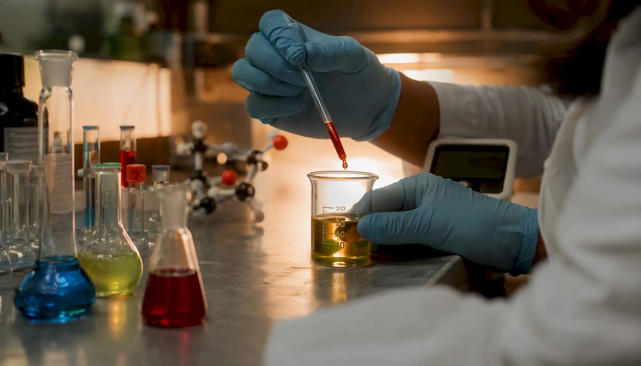
The downstream products are measurable and damaging. Malondialdehyde (MDA) and 4-hydroxynonenal (4-HNE) are the two most clinically tracked reactive carbonyls generated during this cascade. Both form covalent adducts with proteins and DNA, amplifying damage beyond the membrane itself. For white blood cells, membrane disruption means compromised receptor signaling, reduced chemotaxis, and impaired ability to form the tight contacts needed for phagocytosis.
| Damage Type | Biochemical Marker | Functional Consequence |
|---|---|---|
| Lipid peroxidation | MDA, 4-HNE, TBARS | Membrane dysfunction, impaired chemotaxis |
| Protein oxidation | Protein carbonyls | Enzyme inactivation, disrupted signaling |
| DNA damage | 8-OHdG, strand breaks | Apoptosis, mutation risk, impaired proliferation |
| Peroxynitrite attack | Nitrotyrosine | Cytoskeletal disruption, altered cytokine production |
2. Protein oxidation inactivates immune enzymes and disrupts signaling
ROS oxidize amino acid side chains, particularly cysteine, methionine, and tryptophan residues, introducing carbonyl groups and forming disulfide crosslinks that alter protein shape and function. PMC research on ROS signaling distinguishes reversible oxidation, such as sulfenic acid formation on cysteine, from irreversible modifications like sulfonylation or carbonylation, which mark proteins for proteasomal degradation.
In leukocytes, the consequences are specific and consequential. Kinases and phosphatases that regulate cytokine production, phagocytic cup formation, and antigen presentation all depend on precisely folded active sites. Oxidative carbonylation of these enzymes can silence signaling cascades mid-activation, leaving a white blood cell unable to complete the immune response it started. Oxidation of cytoskeletal proteins like actin further impairs the physical mobility neutrophils and macrophages need to reach infection sites.
3. Oxidative DNA damage threatens cell survival and genomic integrity
The hydroxyl radical attacks DNA bases and the deoxyribose backbone with near-diffusion-limited speed. The most common base lesion is 8-hydroxy-2’-deoxyguanosine (8-OHdG), a mutagenic modification that pairs with adenine instead of cytosine during replication. Single- and double-strand breaks follow when the sugar-phosphate backbone is cleaved.
For white blood cells, DNA damage has two immediate consequences. First, it activates p53-dependent apoptosis pathways, shortening leukocyte lifespan and reducing the circulating pool of functional immune cells. Second, in cells that survive with unrepaired lesions, altered gene expression can shift cytokine profiles or impair the precision of adaptive immune responses. Free radical–mediated damage in leukocytes is detectable through assays like the comet assay, which visualizes strand breaks at the single-cell level.
4. The respiratory burst creates self-inflicted leukocyte damage
Here is the central paradox of neutrophil biology: the same oxidative burst that kills bacteria can kill the neutrophil itself. Classic research published in PubMed demonstrated that phagocyte-generated superoxide and hydrogen peroxide can lead to hydroxyl radical formation and premature leukocyte death, and that adding superoxide dismutase or catalase in experimental settings mitigated that self-inflicted damage.
The risk is highest when the phagosomal membrane is leaky, when pathogens are too large to be fully engulfed (frustrated phagocytosis), or when the burst is triggered extracellularly. In those scenarios, HOCl, O2•-, and H2O2 flood the cytoplasm rather than the phagosome. The neutrophil’s own antioxidant reserves, primarily glutathione (GSH) and SOD, are the only barrier between a successful kill and self-destruction.
5. NETosis: a specialized ROS-dependent cell death pathway
Neutrophil extracellular traps (NETs) are web-like structures of decondensed chromatin, histones, and antimicrobial proteins that neutrophils expel to trap and kill pathogens outside the cell. The process, called NETosis, is largely ROS-dependent. Research on neutrophil ROS and NETosis shows that both NOX-dependent and NOX-independent pathways drive the ROS-mediated protein modifications that trigger chromatin decondensation and NET release.
The immune benefit is real: NETs can immobilize bacteria and fungi that evade phagocytosis. The cost is also real. NETosis is a form of cell death, permanently removing that neutrophil from the circulating pool. Excessive or dysregulated NETosis, driven by chronic oxidative stress, contributes to vascular inflammation, thrombosis, and autoimmune tissue damage. It is one of the clearest examples of how free radical white blood cell damage extends beyond the cell itself into systemic pathology.
6. Apoptosis and necrosis: how oxidative stress ends a white blood cell’s life
When oxidative damage crosses a threshold, white blood cells die. The mode of death matters for immune outcomes. Apoptosis, the orderly programmed pathway, produces membrane-enclosed apoptotic bodies that are cleared by macrophages without triggering inflammation. Necrosis, which occurs when damage is too rapid or severe for apoptosis to proceed, releases intracellular contents including DAMPs (damage-associated molecular patterns) that amplify inflammatory signaling and recruit more immune cells.
Oxidative stress causes inflammation partly through this mechanism: necrotic leukocytes spill their oxidized contents into surrounding tissue, activating pattern recognition receptors and sustaining the inflammatory cycle. High-dose ROS exposure tends to push cells toward necrosis rather than apoptosis, which is why uncontrolled oxidative stress in tissues is associated with chronic, low-grade inflammation rather than clean immune resolution.
7. Impaired phagocytosis and antigen presentation reduce pathogen clearance
Oxidative damage does not always kill a white blood cell outright. Often it leaves the cell alive but functionally compromised, a state sometimes called “functional paralysis.” Lipid peroxidation products like 4-HNE modify surface receptors and membrane lipid rafts, reducing the efficiency of pathogen recognition. Protein carbonylation in the actin cytoskeleton impairs the physical mechanics of phagocytic cup formation.
Macrophages and dendritic cells that survive oxidative stress with damaged MHC-II complexes or oxidized antigen-processing enzymes present antigens less effectively to T cells. This breakdown in communication between innate and adaptive immunity means that even when a pathogen is recognized, the signal to mount a targeted adaptive response may be weak or delayed. Oxidation disrupts cellular communication at precisely the molecular interfaces that coordinate these handoffs.
8. Differences in susceptibility across white blood cell types
Not all leukocytes face the same oxidative risk. Neutrophils are the most exposed: they generate the highest ROS loads during respiratory bursts, have relatively short lifespans (hours to days), and rely heavily on GSH and SOD for self-protection. Their high MPO content makes them particularly vulnerable to HOCl-mediated self-damage when the phagosomal seal is incomplete.
Lymphocytes (T cells, B cells, NK cells) are more sensitive to oxidative damage at lower ROS concentrations. They have lower catalase activity than neutrophils and depend more heavily on the thioredoxin system and GSH for redox maintenance. Oxidative damage in lymphocytes tends to manifest as impaired proliferation, altered cytokine secretion, and reduced cytotoxic capacity rather than immediate cell death. Monocytes occupy a middle ground, with moderate antioxidant capacity and a longer lifespan that makes cumulative oxidative damage a particular concern in chronic inflammatory conditions.
9. Chronic oxidative stress links leukocyte damage to systemic disease
Sustained free radical damage to white blood cells does not stay contained at the cellular level. Redox regulation of immune cell function shows that chronic nitro-oxidative stress dysregulates immune metabolism and contributes to disease processes including atherosclerosis and diabetes. In atherosclerosis, oxidatively damaged monocytes and macrophages accumulate in arterial walls, where they take up oxidized LDL and form foam cells, the cellular foundation of plaques.
In diabetes, the mitochondria-NOX crosstalk described earlier shifts persistently toward pro-oxidant states, impairing neutrophil function and increasing infection susceptibility. Autoimmune conditions present a different problem: oxidatively modified self-proteins can become neoantigens, triggering immune responses against the body’s own tissues. The clinical consequences of metabolic oxidative stress span multiple organ systems precisely because leukocytes circulate everywhere.
Key clinical outcomes linked to sustained leukocyte oxidative damage:
- Reduced pathogen clearance and increased infection susceptibility
- Chronic low-grade inflammation from necrotic cell debris and DAMPs
- Accelerated atherosclerosis through foam cell formation
- Impaired glycemic control in diabetes via neutrophil and macrophage dysfunction
- Autoimmune sensitization through oxidatively modified self-antigens
- Lymphocyte depletion and weakened adaptive immune memory
10. Antioxidant defenses inside white blood cells and how they maintain redox balance
White blood cells carry a layered enzymatic defense system specifically calibrated to handle the ROS they generate. Think of it as a relay team, each enzyme passing the oxidative baton to the next until the threat is neutralized.
Superoxide dismutase (SOD) is the first runner. It converts O2•- to H2O2, preventing superoxide from reacting with nitric oxide to form the more damaging peroxynitrite. SOD1 operates in the cytoplasm, SOD2 in the mitochondrial matrix, and SOD3 in the extracellular space. Superoxide dismutase supplements that support this enzymatic step address the earliest and most upstream point in the ROS cascade.
Catalase accepts the H2O2 that SOD produces and converts it to water and oxygen. It is highly concentrated in peroxisomes and provides a high-capacity, non-saturable defense against H2O2 accumulation.
Glutathione (GSH) and glutathione peroxidase (GPx) handle H2O2 and lipid hydroperoxides that catalase misses, particularly in the cytoplasm and mitochondria. GSH is the cell’s most abundant small-molecule antioxidant, and its ratio to oxidized glutathione (GSSG) is a reliable index of cellular redox status.
Thioredoxin (Trx) reduces oxidized cysteine residues on proteins, making it the primary repair enzyme for reversible protein oxidation. It works in tandem with thioredoxin reductase and NADPH.
Nrf2 (nuclear factor erythroid 2-related factor 2) is the master transcription factor that coordinates all of the above. Under oxidative stress, Nrf2 dissociates from its inhibitor Keap1, translocates to the nucleus, and upregulates genes encoding SOD, catalase, GPx, and GSH synthesis enzymes. Nrf2 activity is the primary determinant of how resilient a leukocyte is under sustained oxidative challenge.
| Enzyme / Antioxidant | Primary Function | Cellular Location |
|---|---|---|
| SOD1/SOD2/SOD3 | Converts O2•- to H2O2 | Cytoplasm / mitochondria / extracellular |
| Catalase | Converts H2O2 to H2O + O2 | Peroxisomes |
| Glutathione (GSH) / GPx | Reduces H2O2 and lipid hydroperoxides | Cytoplasm, mitochondria |
| Thioredoxin (Trx) | Repairs reversible protein cysteine oxidation | Cytoplasm, nucleus |
| Nrf2 | Master regulator of antioxidant gene expression | Nucleus (activated) |
Pro Tip: Targeted enzymatic support, particularly at the SOD step, addresses the upstream source of the ROS cascade rather than mopping up downstream damage. Indiscriminate high-dose antioxidant supplementation can blunt the low-level ROS signaling that lymphocytes need for activation and that neutrophils need to initiate a proper burst. The goal is redox balance, not zero ROS.
How researchers detect oxidative damage in blood cells
Measuring free radical white blood cell damage requires choosing the right assay for the right question. Each method captures a different slice of the oxidative picture, and each carries interpretive caveats.
- MDA and TBARS (thiobarbituric acid reactive substances): MDA is the most widely used lipid peroxidation marker; TBARS is the colorimetric assay that quantifies it. Sensitive and inexpensive, but prone to artifactual oxidation during sample processing if not handled carefully.
- 4-HNE: more specific than MDA for lipid peroxidation; forms stable protein adducts that can be detected by ELISA or immunohistochemistry, making it useful for cell-specific localization.
- Protein carbonyls: measured by DNPH derivatization; a direct index of irreversible protein oxidation. Elevated carbonyls in isolated leukocytes indicate that the antioxidant system has been overwhelmed.
- EPR (electron paramagnetic resonance) spin-trapping: the gold standard for directly detecting radical species in real time. Spin-trap compounds like PBN react with short-lived radicals to form stable adducts detectable by EPR. Research with PBN showed that spin-trapping compounds extended polymorphonuclear leukocyte lifespan after respiratory-burst-induced superoxide generation, suggesting a protective effect of targeted radical scavengers.
- Immunospin trapping: combines spin-trapping with antibody detection, allowing protein-centered radicals to be localized to specific cellular proteins.
- Flow cytometry with oxidation-sensitive dyes: probes like DCFH-DA or MitoSOX Red allow real-time ROS measurement in live, individual leukocytes, enabling cell-type-specific analysis.
- Comet assay: single-cell gel electrophoresis that visualizes DNA strand breaks; particularly useful for lymphocyte oxidative DNA damage studies.
Pro Tip: Plasma MDA or TBARS measurements give a systemic oxidative stress signal but cannot tell you which cell type is damaged or whether the damage is occurring in circulating leukocytes specifically. For leukocyte-specific conclusions, isolate the cell population first, then run the assay on the cell lysate or use flow cytometry on intact cells. Sample handling time and temperature are critical variables; oxidation continues ex vivo.
Can free radical damage to white blood cells be reversed?
Some oxidative modifications are reversible; others are not. Reversible changes, such as sulfenic acid formation on cysteine residues or methionine sulfoxidation, can be repaired by thioredoxin and methionine sulfoxide reductase without replacing the protein. Irreversible adducts, including carbonylated proteins and MDA-DNA crosslinks, require proteasomal degradation of the damaged molecule and synthesis of a replacement, which depends on cell turnover.
The practical implication: early-stage oxidative stress is more recoverable than chronic, accumulated damage. Strategies with the strongest mechanistic rationale include:
- Reduce exposures: cigarette smoke, air pollutants, and ionizing radiation are the most potent exogenous ROS sources. Reducing exposure lowers the baseline oxidative load on circulating leukocytes.
- Optimize metabolic control: in diabetes and metabolic syndrome, the mitochondria-NOX crosstalk drives persistent pro-oxidant states. Glycemic control directly reduces leukocyte ROS generation.
- Dietary polyphenols: compounds like quercetin, resveratrol, and curcumin activate Nrf2 and upregulate endogenous antioxidant enzymes. The National Cancer Institute notes that dietary antioxidants can neutralize free radicals, but their relationship to disease risk is complex and context-dependent.
- Targeted enzymatic antioxidant support: SOD mimetics and bioavailable SOD formulations address the upstream step in the ROS cascade. Targeted antioxidant strategies that support specific enzymatic pathways have a stronger mechanistic rationale than broad-spectrum antioxidant loading.
- Glutathione support: N-acetylcysteine (NAC) is the most studied GSH precursor; it replenishes intracellular GSH and has been used in clinical settings to reduce oxidative burden in inflammatory conditions.
The limits of indiscriminate supplementation deserve emphasis. High-dose vitamin C or vitamin E given at the wrong time can blunt the ROS signaling that lymphocytes need for activation and that neutrophils need to mount an effective burst. Research on reducing oxidative stress for immune function reinforces that timing and targeting matter as much as the antioxidant chosen.
Pro Tip: Clinical trials on antioxidant supplementation have produced mixed results partly because most trials use non-targeted, high-dose approaches that interfere with physiological ROS signaling. Trials using enzymatic antioxidants or Nrf2 activators at doses that restore rather than suppress redox balance show more consistent benefit.
What current research shows about targeted antioxidant strategies
The mechanistic picture of leukocyte oxidative damage is well-established. The therapeutic translation is still catching up. Several directions show genuine promise, though the evidence base varies.
- NOX inhibitors: compounds targeting NOX2 specifically are under investigation for conditions where excessive respiratory burst drives tissue damage, including sepsis and chronic granulomatous disease. Selectivity remains a challenge; broad NOX inhibition risks impairing the antimicrobial burst entirely.
- Nrf2 modulators: sulforaphane (from cruciferous vegetables) and other electrophilic compounds activate Nrf2 by modifying Keap1 cysteines. Small clinical trials show increases in leukocyte GSH and reduced oxidative damage markers, though large randomized trials are limited.
- SOD and catalase systems: bioavailable SOD formulations have shown reductions in systemic antioxidant markers in preliminary studies. The mechanistic rationale is strong given SOD’s upstream position in the ROS cascade.
- Mitochondria-targeted antioxidants: compounds like MitoQ, which concentrate in the mitochondrial matrix, address the mitochondrial ROS source directly. Early data in metabolic disease models are encouraging; human trial data remain limited.
The central challenge in translating antioxidant research into clinical benefit is that ROS are not uniformly harmful. Research on ROS roles in lymphocyte activation confirms that NADPH oxidase and mitochondrial ROS are critical for lymphocyte activation and development. Interventions that eliminate ROS rather than restore balance can suppress the immune responses they are meant to protect. The therapeutic goal is a narrower target than it first appears: reduce pathological ROS without silencing physiological signaling.
Free Radicals, Antioxidants in Disease and Health summarizes that at high concentrations ROS cause lipid peroxidation, protein oxidation, and DNA lesions implicated in chronic disease, while endogenous and exogenous antioxidants act as scavengers. The clinical challenge is that the line between therapeutic scavenging and immunosuppressive interference is concentration-dependent and cell-type-specific.
The nuance most antioxidant conversations miss
The science of free radical white blood cell damage is genuinely complex, and the popular conversation around antioxidants tends to flatten it into a simple good-versus-evil story. What the research actually shows is more interesting and more demanding.
Targeted enzymatic support, particularly at the SOD and glutathione steps, addresses the upstream architecture of the ROS cascade rather than applying a broad chemical blanket. The timing of any intervention relative to immune activation matters. A supplement that reduces oxidative burden during chronic low-grade inflammation is doing something categorically different from one taken during an acute infection, when the respiratory burst is exactly what you want your neutrophils to be doing.
More clinical trials with leukocyte-specific endpoints are needed. Most existing antioxidant trials measure plasma markers, not cell-type-specific oxidative damage in isolated neutrophils or lymphocytes. Until that gap closes, the most defensible approach is to address modifiable exposures, support metabolic health, and use targeted enzymatic strategies rather than high-dose broad-spectrum supplementation. The goal is a well-regulated immune system, not a chemically suppressed one.
Targeted enzymatic antioxidant support for white blood cell health
If the research on free radical white blood cell damage points toward one practical direction, it is this: targeted enzymatic support at the SOD step, combined with lifestyle strategies that reduce the underlying oxidative load, is a more defensible approach than generic antioxidant megadosing.
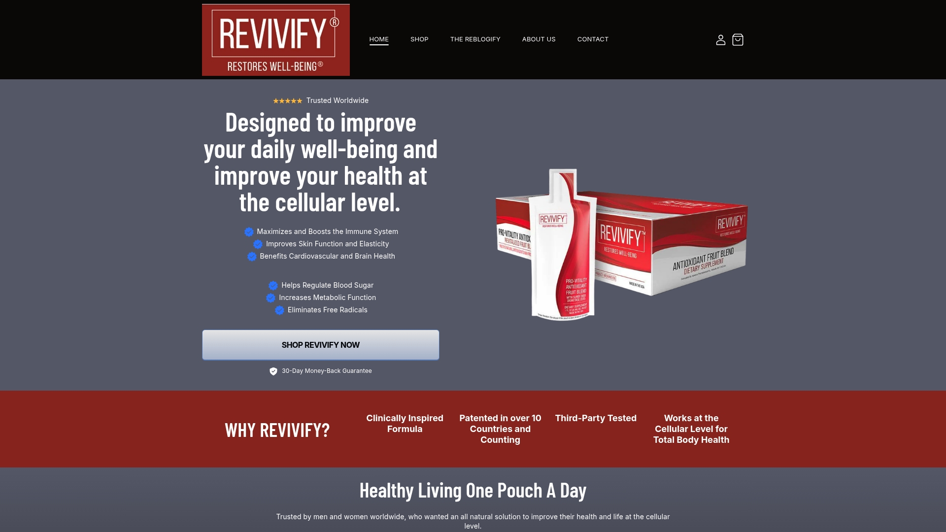
Tryrevivify’s Revivify is a daily supplement in gel form that combines superoxide dismutase, prebiotic fiber, polyphenols, and lactobacillus in a patented formula designed to support your body’s antioxidant defenses at the cellular level. It is third-party tested and plant-based, with subscription options and a risk-free guarantee. SOD addresses the upstream step in the ROS cascade, before H2O2 accumulates and before hydroxyl radical formation becomes a threat to your leukocyte membranes and DNA. Before starting any new supplement, speak with your clinician, particularly if you have a chronic condition or are managing medications that affect immune function. When you are ready to take a targeted approach, visit Tryrevivify to learn more about Revivify and find the option that fits your health goals.
Sources
The following peer-reviewed reviews and clinical overviews formed the evidence base for this article. Consult primary studies before making any changes to clinical care.
- Oxidative Stress: Causes, Symptoms & Treatment
- Free radicals and inflammation. Protection of phagocytosine leukocytes by superoxide dismutase
- ROS signaling in innate immunity via oxidative protein modifications - PMC
- Cancer
This article provides general scientific information and is not a substitute for professional medical advice. Consult a qualified healthcare provider before changing any supplement regimen or clinical care plan.