

How Antioxidants Prevent Arterial Oxidation and Protect Your Heart
Antioxidants prevent arterial oxidation primarily by scavenging reactive oxygen species (ROS) in the subendothelial space, the thin layer beneath the arterial lining where LDL cholesterol is most vulnerable to oxidative attack. When ROS concentrations exceed the artery’s natural defenses, they chemically modify LDL particles, transforming them into oxidized LDL (oxLDL). Macrophages engulf oxLDL without restraint, swelling into foam cells that accumulate into fatty plaques, the structural foundation of atherosclerosis. Antioxidants interrupt this chain at multiple points, not just by neutralizing free radicals, but by preserving the enzymatic machinery that keeps arterial walls functional.
The biochemical term for this process is oxidative stress, a state in which ROS production outpaces the body’s antioxidant defenses. Understanding how antioxidants prevent arterial oxidation requires looking at both the molecular targets they protect and the enzymatic systems they support.
Key antioxidant actions in the arterial wall:
- Scavenging superoxide radicals and hydrogen peroxide before they oxidize LDL
- Preserving nitric oxide (NO) bioavailability by preventing eNOS uncoupling
- Maintaining the glutathione (GSH) redox ratio to protect critical cysteine residues in enzymes
- Activating protective pathways such as heme oxygenase-1 (HO-1) induction
- Inhibiting NADPH oxidase activity to reduce ROS generation at the source
- Chelating transition metal ions that catalyze lipid peroxidation in arterial cells
- Activating Nrf2 transcription to upregulate endogenous antioxidant gene expression
Pro Tip: Think of your arterial antioxidant system as a relay team. Superoxide dismutase (SOD) handles the first leg by converting superoxide into hydrogen peroxide, then glutathione peroxidase and catalase take the baton to neutralize that hydrogen peroxide before it causes further damage. The whole team has to show up for the race to be won.

How oxidative stress drives atherosclerosis at the molecular level
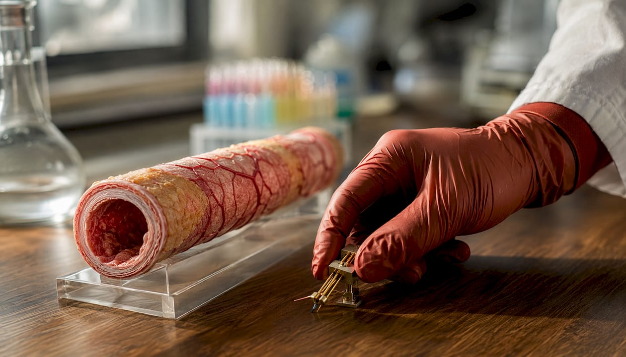
Atherosclerosis is not simply a plumbing problem of cholesterol buildup. It is a chronic inflammatory disease, and oxidative stress is one of its earliest and most persistent drivers. The arterial wall generates ROS from several enzymatic sources, each contributing to a redox environment that, when uncontrolled, progressively damages lipids, proteins, and the endothelial cells that line every blood vessel in your body.
The two primary ROS-generating enzymes in the arterial wall are NADPH oxidase and uncoupled endothelial nitric oxide synthase (eNOS). NADPH oxidase produces superoxide radicals directly in vascular cells, including endothelial cells, smooth muscle cells, and macrophages. Under normal conditions, eNOS produces NO, a vasodilator that keeps arteries relaxed and prevents platelet aggregation. When eNOS loses its cofactor tetrahydrobiopterin (BH4) or undergoes S-glutathionylation of its cysteine residues, it “uncouples,” switching from NO production to superoxide generation. The same enzyme that protects the artery becomes a source of the very damage it was meant to prevent.
Oxidative stress in arteries generates ROS from these endothelial and immune cells, and the downstream consequences are severe. Superoxide reacts with NO to form peroxynitrite, a potent oxidant that further depletes BH4 and accelerates eNOS uncoupling in a self-reinforcing cycle. LDL particles trapped in the subendothelial space are oxidized by these ROS, and the resulting oxLDL triggers macrophage infiltration, foam cell formation, and inflammatory cytokine release.
| ROS Type | Primary Source | Arterial Effect |
|---|---|---|
| Superoxide (O₂•⁻) | NADPH oxidase, uncoupled eNOS | Oxidizes LDL, depletes NO, promotes inflammation |
| Hydrogen peroxide (H₂O₂) | SOD-mediated superoxide dismutation | Activates redox-sensitive transcription factors; can be cytotoxic at high levels |
| Peroxynitrite (ONOO⁻) | Reaction of superoxide with NO | Nitrosylates proteins, oxidizes BH4, impairs eNOS function |
| Hydroxyl radical (•OH) | Fenton reaction with iron/copper | Directly damages DNA, lipids, and proteins in arterial cells |
| Hypochlorous acid (HOCl) | Myeloperoxidase in macrophages | Oxidizes LDL in plaques, promotes foam cell formation |
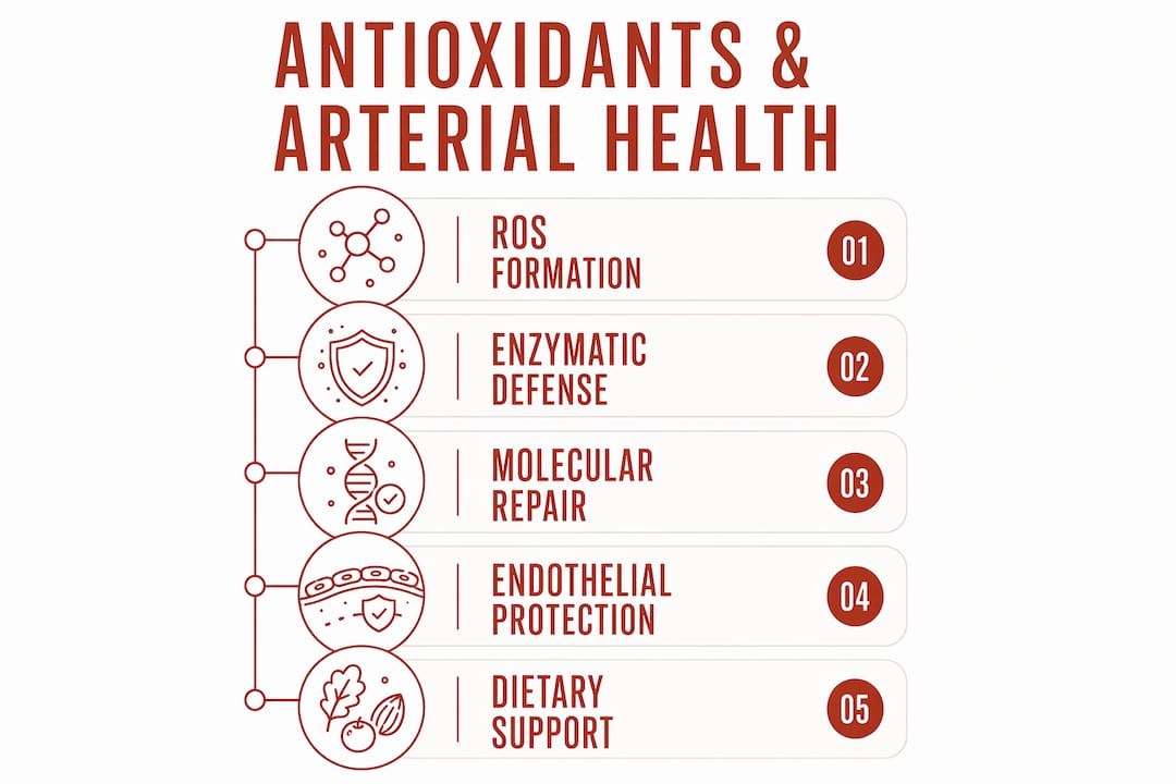
Endothelial dysfunction is the clinical consequence of this oxidative cascade. When NO bioavailability drops, arteries lose their ability to dilate in response to blood flow, platelet adhesion increases, and the expression of adhesion molecules like VCAM-1 and ICAM-1 rises, recruiting more monocytes into the arterial wall. This is the point at which endothelial oxidative damage transitions from a biochemical event to a clinically measurable disease process.
Flavonoids and polyphenols can inhibit NADPH oxidase activity directly, reducing superoxide production at the source rather than simply mopping up radicals after the fact. This upstream intervention is one reason dietary antioxidant patterns show stronger vascular protection than isolated radical-scavenging supplements.
How antioxidants preserve endothelial function at the molecular level
The most clinically consequential way antioxidants protect arteries is not by neutralizing a free radical in isolation. It is by preserving the function of eNOS, the enzyme responsible for producing NO in the endothelium. When the redox environment in the arterial wall tips toward oxidation, eNOS uncouples, and NO production collapses. Antioxidants prevent this by maintaining the molecular conditions eNOS needs to stay functional.
The eNOS coupling mechanism and why it matters
eNOS uncoupling occurs through two primary routes. First, oxidative depletion of BH4, a redox-sensitive cofactor that stabilizes the enzyme’s active state. Second, S-glutathionylation of critical cysteine residues at positions 689 and 908 in the eNOS protein, a modification driven by a low glutathione (GSH) to oxidized glutathione (GSSG) ratio. Antioxidants maintain the GSH:GSSG ratio by scavenging the ROS that would otherwise oxidize glutathione, keeping those cysteine residues in their reduced, functional state. Vitamin C stabilizes BH4 levels directly, providing an additional layer of protection against eNOS uncoupling.
Enzymatic antioxidants: SOD, glutathione, and their isoforms
Superoxide dismutase (SOD) is the first line of enzymatic defense against superoxide in the arterial wall. Think of it as the first runner in a relay team: it converts superoxide into hydrogen peroxide, passing the baton to downstream enzymes. Three isoforms operate in different cellular compartments:
- SOD1 (Cu/Zn-SOD): Located in the cytoplasm and on the inner mitochondrial membrane
- SOD2 (Mn-SOD): Found in the mitochondrial matrix, where ROS production from electron transport is highest
- SOD3 (EC-SOD): Extracellular, positioned precisely where LDL oxidation in the subendothelial space occurs
Critically, SOD does not work alone. In animal models, overexpression of SOD1 without sufficient catalase to process the resulting hydrogen peroxide actually enhanced atherosclerosis development, because hydrogen peroxide itself is a substrate for further oxidative reactions. The relay only works when every runner shows up.
Glutathione (GSH) operates as both a direct antioxidant and a cofactor for glutathione peroxidase, the enzyme that reduces hydrogen peroxide and lipid hydroperoxides to harmless products. Dietary antioxidants preserve vascular function in part by maintaining a high GSH:GSSG ratio, which keeps glutathione peroxidase active and eNOS coupled.
Heme oxygenase-1 induction: the pathway beyond radical scavenging
One of the most underappreciated mechanisms of vascular antioxidant protection involves heme oxygenase-1 (HO-1), an enzyme induced in vascular cells in response to oxidative stress. HO-1 degrades heme into biliverdin, carbon monoxide, and free iron, producing products with anti-inflammatory and cytoprotective properties. Research on probucol, a redox-active compound, demonstrated that HO-1 induction by antioxidants mediates vascular protection through cell-specific effects: inhibiting macrophage accumulation, stimulating reendothelialization, and blocking smooth muscle cell proliferation. Strikingly, this protection required the sulfur atoms in probucol’s structure, not its phenolic groups, suggesting that 2-electron redox reactions involving cysteine residues in key enzymes are more central to atherogenesis than simple radical scavenging.
Key antioxidant effects on endothelial function:
- Maintaining BH4 levels to keep eNOS coupled and producing NO
- Preserving the GSH:GSSG ratio to prevent S-glutathionylation of eNOS cysteine residues
- Inducing HO-1 to provide anti-inflammatory and cytoprotective effects in vascular cells
- Activating Nrf2, a transcription factor that upregulates endogenous antioxidant gene expression
- Inhibiting VCAM-1 and ICAM-1 expression to reduce monocyte recruitment into the arterial wall
- Protecting NO from oxidation by peroxynitrite, preserving vasodilatory capacity
Pro Tip: Vitamin C and dietary polyphenols work as a team here. Vitamin C stabilizes BH4 and recycles vitamin E after it neutralizes a lipid radical, while polyphenols like quercetin and catechins inhibit NADPH oxidase upstream. No single molecule does all of this alone, which is exactly why food beats a pill.
Flavonoids and polyphenols also activate Nrf2, a master transcription factor that switches on a battery of endogenous antioxidant genes including those encoding SOD, glutathione peroxidase, and HO-1. This means dietary antioxidants do not just neutralize ROS directly; they strengthen your body’s own defenses at the genetic level, producing a sustained protective effect that outlasts the presence of any individual molecule.
What the clinical evidence actually shows about antioxidants and cardiovascular risk
The clinical picture on antioxidants and cardiovascular disease is more nuanced than either enthusiasts or skeptics typically acknowledge. Observational data and randomized controlled trials (RCTs) tell different stories, and understanding why requires looking carefully at what each type of study actually measures.
What observational studies show
Large-scale epidemiological research consistently links antioxidant-rich dietary patterns to lower cardiovascular disease (CVD) risk. A review of 95 observational studies involving more than 2 million participants found that dietary patterns high in whole-food antioxidants correlate with lower cardiovascular risk. Plant-based diets rich in fruits, vegetables, legumes, and whole grains are associated with reduced rates of coronary heart disease, stroke, and cardiovascular mortality across diverse populations. The Harvard Nurses’ Health Study and Health Professionals Follow-Up Study both found inverse associations between dietary antioxidant intake and CVD incidence, though these associations were strongest for food-derived antioxidants rather than supplements.
Where randomized controlled trials diverge
RCTs on isolated antioxidant supplements have largely failed to replicate the protective effects seen in observational studies. Trials of vitamin E, beta-carotene, and vitamin C as standalone supplements have not demonstrated significant reductions in cardiovascular events, and some have raised safety concerns.
| Study Type | Population | Antioxidant Tested | Outcome | Key Limitation |
|---|---|---|---|---|
| Observational (prospective cohort) | General population, dietary patterns | Whole-food antioxidant intake | Lower CVD risk | Cannot establish causation; confounding by overall diet quality |
| RCT (HOPE trial) | High-risk CVD patients | Vitamin E supplement | No cardiovascular benefit | Single antioxidant; advanced disease at enrollment |
| RCT (ATBC trial) | Male smokers | Beta-carotene supplement | Increased lung cancer risk | High-risk population; pharmacological doses |
| RCT (PREDIMED) | High CVD risk adults | Mediterranean diet (whole foods) | Reduced major CVD events | Diet-based, not isolated supplement |
| Meta-analysis | Multiple populations | Mixed antioxidant supplements | Inconsistent results | Heterogeneous populations, doses, and endpoints |
The PREDIMED trial stands out because it tested a whole-food dietary pattern rich in antioxidants rather than an isolated supplement. Participants assigned to a Mediterranean diet supplemented with extra-virgin olive oil or nuts showed reduced rates of major cardiovascular events compared to a control diet. This result aligns with the observational data and points toward dietary pattern rather than any single molecule as the operative variable.
Observational studies show a protective effect of antioxidant-rich diets, while clinical trials on supplements often yield disappointing results for cardiovascular outcomes. The gap between these two bodies of evidence is not a contradiction. It is a signal about what antioxidants actually need to work.
Why antioxidant clinical trials so often miss the mark
The failure of antioxidant supplement trials to show cardiovascular benefit is one of the more instructive puzzles in modern nutrition science. The biochemical rationale is sound, the observational data is consistent, yet trial after trial comes up empty. Several biological and methodological factors explain this gap.
ROS are not purely harmful
Physiological ROS levels function as signaling molecules in vascular cells. Superoxide and hydrogen peroxide at low concentrations regulate vascular tone, promote endothelial repair, and modulate immune responses. Indiscriminate antioxidant suppression can disrupt these normal signaling functions, potentially offsetting the benefits of reducing oxidative damage. High-dose antioxidant supplements do not distinguish between harmful excess ROS and the physiological ROS that cells need for normal function. This is a fundamental biological problem that no trial design can fully overcome when the intervention is a pharmacological dose of a single molecule.
The synergy problem
Antioxidants do not work in isolation in the body. They operate as an interconnected network in which each molecule depends on others for regeneration and activity. After vitamin C donates an electron to neutralize a free radical, it becomes a dehydroascorbate radical. Phytochemicals such as hesperetin, found in citrus fruits, regenerate vitamin C back to its active form. Vitamin E is recycled by vitamin C after quenching lipid radicals in LDL particles. Glutathione regenerates other antioxidants through the glutaredoxin system. A supplement containing only vitamin E or only vitamin C extracts one player from this network and expects it to perform without its teammates.
Bioavailability and timing
Most antioxidant trials enroll participants who already have established cardiovascular disease or significant atherosclerotic burden. At that stage, oxidative damage has been accumulating for decades. Giving a supplement to someone with advanced plaques is analogous to applying sunscreen after a sunburn. The protective window for antioxidant intervention is likely earlier in the disease process, during the phase when LDL oxidation and endothelial dysfunction are just beginning.
Bioavailability compounds this problem. Many polyphenols are poorly absorbed from supplements compared to their food-matrix forms, where fiber, cofactors, and other phytochemicals enhance uptake. The dose that reaches the arterial wall from a capsule is often far lower than what a food-based intake delivers over time.
Additional factors that undermine antioxidant trial results:
- Failure to stratify participants by baseline oxidative stress status, meaning some participants may not have had elevated ROS to begin with
- Short trial durations that cannot capture the decades-long process of atherosclerosis development
- Use of synthetic antioxidant forms (e.g., dl-alpha-tocopherol) with lower biological activity than natural food forms
- Lack of assessment of antioxidant network status, so researchers cannot tell whether the supplement actually changed the redox environment
- Potential pro-oxidant effects of high-dose supplements in specific populations, particularly smokers and those with existing disease
The lesson from these failures is not that antioxidants do not matter for arterial health. It is that they matter most when delivered as part of a complex dietary pattern, consistently, over time, before disease is established. Past clinical trial failures reflect a misunderstanding of antioxidants as post-hoc extinguishers rather than ongoing regulatory agents requiring consistent presence through diet.
How dietary antioxidants deliver what supplements cannot
The gap between supplement trials and observational diet studies is not a statistical artifact. It reflects a genuine biological difference between eating antioxidants as part of a whole food and swallowing them in a capsule. Antioxidant-rich diets reflect complex nutrient networks providing fiber, micronutrients, and synergistic phytochemicals, and that complexity is precisely what explains the benefits that supplements cannot replicate.
Redox balance and the eNOS connection
A diet consistently high in antioxidant-rich foods maintains the GSH:GSSG ratio at levels that keep eNOS coupled and producing NO. This is not a one-time event. It is a continuous biochemical condition that requires a steady supply of reducing equivalents from food. Dietary flavonoids and polyphenols, found in green tea, cocoa, berries, and extra-virgin olive oil, have been shown in animal models to increase BH4 levels and NO bioavailability, directly alleviating eNOS uncoupling and reducing oxidative stress in the vascular wall. No supplement trial has replicated this effect at scale, partly because no capsule delivers the full polyphenol profile of a cup of green tea.
The antioxidant regeneration network in food
Whole foods contain the complete regeneration network that isolated supplements lack. Vitamin C in an orange comes packaged with hesperetin and other flavonoids that recycle it after it neutralizes a radical. Vitamin E in almonds arrives with selenium and other cofactors that support glutathione peroxidase activity. This co-packaging is not incidental. It is the result of plants evolving their own antioxidant defense systems, which happen to map remarkably well onto human vascular biochemistry.
Dietary antioxidant sources with documented vascular benefits:
- Flavonoids (green tea, cocoa, berries, onions): Inhibit NADPH oxidase, protect LDL from oxidation, activate Nrf2
- Polyphenols (extra-virgin olive oil, red wine, pomegranate): Increase eNOS expression and NO bioavailability, reduce VCAM-1 expression
- Vitamin C (citrus, bell peppers, kiwi): Stabilizes BH4, recycles vitamin E, scavenges superoxide and hydroxyl radicals
- Vitamin E (nuts, seeds, wheat germ): Protects LDL lipid core from peroxidation, prevents foam cell formation
- Carotenoids (tomatoes, kale, carrots): Quench singlet oxygen, reduce lipid peroxidation in arterial cells
- Glutathione precursors (cruciferous vegetables, garlic, onions): Support GSH synthesis and the glutathione redox system
Why enzymatic antioxidants like SOD matter for supplementation strategy
At Tryrevivify, we’ve built our formula around a recognition that the enzymatic antioxidant system is the body’s primary defense against arterial oxidation, not dietary vitamins alone. SOD is the rate-limiting enzyme in superoxide clearance, and its activity in the vascular wall directly determines how much superoxide is available to oxidize LDL and deplete NO. Tryrevivify combines superoxide dismutase with prebiotic fiber in a patented formula designed to support this enzymatic defense at the cellular level, addressing the root of arterial oxidation rather than its downstream consequences.
The prebiotic fiber component matters here too. Gut microbiome composition influences systemic antioxidant markers, including circulating glutathione and inflammatory cytokines that modulate vascular oxidative stress. A healthy gut microbiome produces short-chain fatty acids that reduce intestinal permeability and systemic inflammation, two factors that feed directly into the oxidative burden on arterial walls.
Pro Tip: When building a diet for vascular antioxidant protection, prioritize variety over volume. A handful of blueberries, a cup of green tea, a tablespoon of extra-virgin olive oil, and a serving of cruciferous vegetables each day covers multiple nodes of the antioxidant network simultaneously. No single superfood does what the combination does.
The evidence from observational studies, mechanistic research, and the selective successes of diet-based trials converges on one conclusion: the most effective way to maintain antioxidant protection in your arteries is through a consistent, varied, whole-food dietary pattern that keeps the entire redox network active. Supplements can play a supporting role, particularly when they target enzymatic pathways like SOD that diet alone may not fully saturate, but they work best as additions to a strong dietary foundation, not replacements for one.
Understanding the role of antioxidants in healing extends well beyond the arterial wall, touching cellular repair processes throughout the body that share the same enzymatic machinery. The vascular benefits of a well-maintained antioxidant system are, in that sense, one expression of a broader cellular resilience that whole-food nutrition builds over time.
Key Takeaways
Antioxidants prevent arterial oxidation by maintaining the enzymatic and molecular conditions that keep LDL intact, eNOS coupled, and NO bioavailable, and this protection works best when delivered through consistent whole-food dietary patterns rather than isolated supplements.
| Point | Details |
|---|---|
| ROS drive atherosclerosis | NADPH oxidase and uncoupled eNOS generate superoxide that oxidizes LDL and depletes NO, initiating foam cell formation and plaque development. |
| eNOS coupling is the key target | Antioxidants preserve the GSH:GSSG ratio and BH4 levels to keep eNOS producing NO rather than superoxide, protecting vascular dilation. |
| SOD leads the enzymatic defense | Superoxide dismutase converts superoxide to hydrogen peroxide, but requires downstream enzymes like glutathione peroxidase and catalase to complete the detoxification relay. |
| Diet outperforms supplements | A review of 95 observational studies with more than 2 million participants found whole-food antioxidant patterns correlate with lower cardiovascular risk, while isolated supplement trials have largely failed to show benefit. |
| HO-1 induction extends protection | Antioxidants that activate heme oxygenase-1 provide vascular protection beyond radical scavenging, inhibiting macrophage accumulation and promoting reendothelialization. |