

How Inflammation Triggers Insulin Resistance: 2026 Guide
Insulin resistance is defined as the failure of cells in your liver, muscle, and fat tissue to respond normally to insulin, causing blood sugar to rise unchecked. Understanding how inflammation triggers insulin resistance is the key to grasping why so many people develop type 2 diabetes without obvious warning signs. Chronic low-grade inflammation shifts immune cells called macrophages into a pro-inflammatory state, flooding tissues with cytokines like TNF-α and IL-6 that block insulin receptor signaling. This process, known clinically as “meta-inflammation,” operates silently for years before a diagnosis appears. Recognizing the inflammation and insulin resistance link is the first step toward meaningful prevention.
How does inflammation trigger insulin resistance at the molecular level?
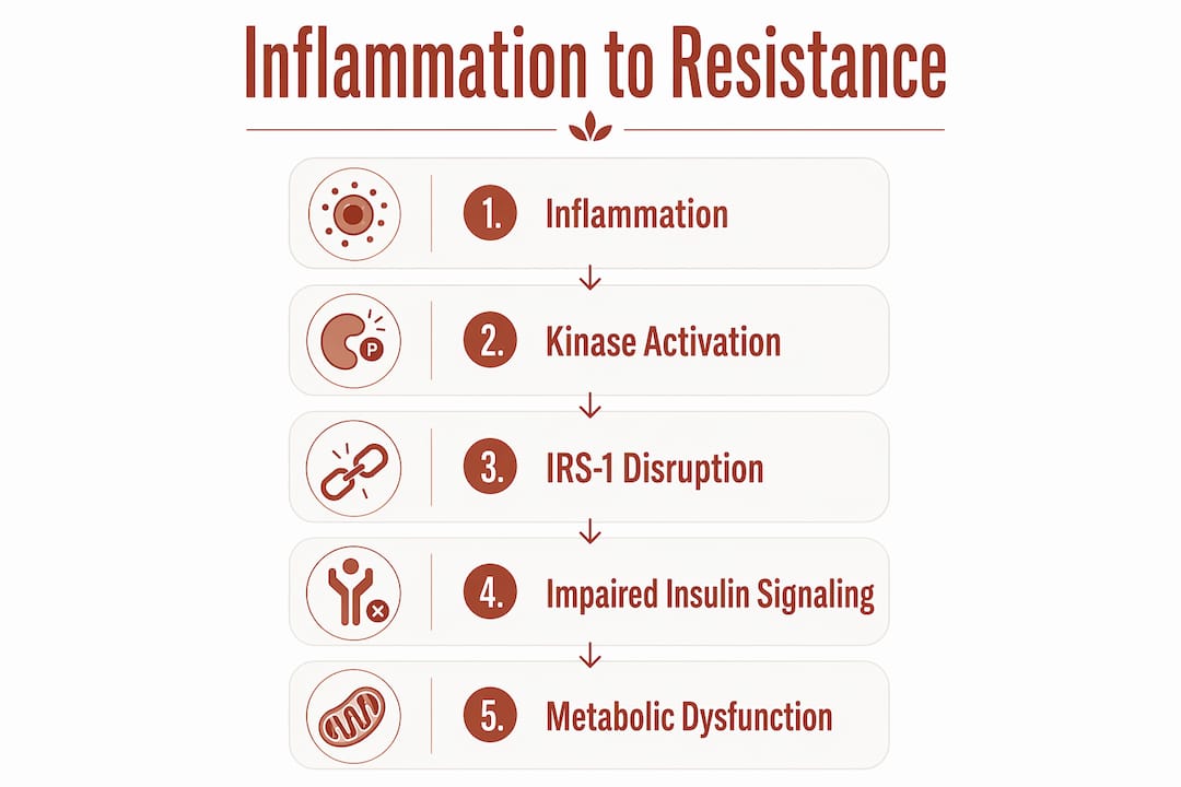
Inflammation disrupts insulin signaling through a precise chain of molecular events. The central target is insulin receptor substrate-1, or IRS-1, a protein that acts as the first relay switch after insulin binds to its receptor on a cell. When IRS-1 works correctly, it activates a cascade that tells the cell to absorb glucose from the blood. When inflammation interferes, that switch gets jammed.
The jamming mechanism involves two inflammatory kinases: IKKβ and JNK. Both enzymes are activated by pro-inflammatory signals. Once active, they phosphorylate IRS-1 at the wrong sites, which is like pressing the wrong key on a keyboard. The signal gets garbled, and glucose uptake stalls. NF-κB pathway activation compounds this problem by suppressing the proteins needed for insulin sensitivity in liver and fat cells, creating a broader signaling failure.

Cytokines play a starring role in this process. TNF-α and IL-6, both released by pro-inflammatory macrophages, directly impair IRS-1 function. A third group of proteins called SOCS (suppressor of cytokine signaling) proteins adds another layer of interference by tagging IRS-1 for degradation. Less IRS-1 means less insulin signaling capacity, even when insulin levels in the blood are high.
The macrophage shift is the trigger that starts this entire sequence. Healthy adipose tissue contains anti-inflammatory M2 macrophages that support tissue repair. Macrophage polarization from M2 to M1 leads to secretion of TNF-α and IL-6, converting fat tissue from a metabolic support system into an inflammatory engine. This shift is the cellular origin of insulin resistance in obesity.
Key molecular players in the inflammation-insulin resistance pathway:
- IRS-1 phosphorylation disruption: IKKβ and JNK block the insulin signal at its first relay point.
- NF-κB activation: This transcription factor suppresses insulin-sensitizing proteins across liver and fat cells.
- TNF-α and IL-6: Pro-inflammatory cytokines secreted by M1 macrophages that directly impair IRS-1 function.
- SOCS proteins: Accelerate IRS-1 degradation, reducing the cell’s total capacity to respond to insulin.
- M1 macrophage dominance: Shifts adipose tissue from an anti-inflammatory to a pro-inflammatory environment.
Pro Tip: Tracking high-sensitivity C-reactive protein (hs-CRP) in routine bloodwork gives you a practical window into systemic inflammation levels before insulin resistance becomes clinically apparent.
Which risk factors increase inflammation-driven insulin resistance?
Several lifestyle and physiological conditions amplify the inflammatory signals that impair insulin sensitivity. Knowing these factors helps you identify where your own metabolic risk is highest.
-
Visceral fat accumulation. Visceral fat acts as an endocrine organ, secreting TNF-α and IL-6 directly into the portal circulation, which feeds straight into the liver. This makes abdominal fat uniquely damaging compared to subcutaneous fat stored under the skin. As fat cells enlarge (a process called adipose tissue hypertrophy), they become hypoxic and begin to die, recruiting waves of pro-inflammatory macrophages that worsen the cycle.
-
Physical inactivity. Muscle tissue is the body’s largest glucose sink. When you don’t use your muscles regularly, they become less sensitive to insulin and contribute less to clearing blood sugar. Inactivity also allows visceral fat to accumulate, compounding the inflammatory burden.
-
Chronic stress. Sustained psychological stress elevates cortisol, which promotes fat storage in the abdomen and activates inflammatory pathways. Cortisol also suppresses anti-inflammatory immune responses, leaving pro-inflammatory signals less regulated.
-
Sleep deprivation. Poor sleep quality raises inflammatory markers and impairs glucose metabolism within days. Even one week of restricted sleep measurably increases insulin resistance in otherwise healthy adults.
-
Nutrient overload and obesity. Meta-inflammation is a chronic maladaptive immune response to nutrient overload that creates a self-sustaining cycle worsening insulin resistance over time. Unlike acute inflammation that resolves after an injury, meta-inflammation persists as long as excess nutrients keep activating immune sensors in fat and liver tissue.
-
Oxidative and endoplasmic reticulum stress. Oxidative stress generates reactive oxygen species that activate the same inflammatory kinases (IKKβ and JNK) that block IRS-1. Endoplasmic reticulum stress, triggered by protein misfolding from nutrient overload, independently activates JNK and NF-κB, adding a second inflammatory input.
How does the inflammation-insulin resistance cycle affect metabolic health?
The relationship between inflammation and insulin resistance is bidirectional. Insulin resistance exacerbates inflammation through cellular stress pathways, which then further impairs insulin sensitivity. This feedback loop is what makes metabolic disease so difficult to reverse once it takes hold.
The consequences spread across three key tissues. In the liver, impaired insulin signaling causes uncontrolled glucose production even when blood sugar is already elevated. In skeletal muscle, reduced glucose uptake means the body’s largest energy consumer stops clearing sugar from the blood efficiently. In adipose tissue, dysfunctional insulin signaling promotes further fat breakdown and cytokine release, feeding more inflammation back into the system.
| Tissue | Effect of impaired insulin signaling | Metabolic consequence |
|---|---|---|
| Liver | Uncontrolled glucose output | Elevated fasting blood sugar |
| Skeletal muscle | Reduced glucose uptake | Postmeal blood sugar spikes |
| Adipose tissue | Increased cytokine secretion | Sustained systemic inflammation |
| Pancreatic beta cells | Compensatory overproduction of insulin | Beta cell exhaustion over time |
The progression toward type 2 diabetes follows a predictable path. Early insulin resistance forces the pancreas to produce more insulin to maintain normal blood sugar. Over years, beta cells exhaust themselves trying to compensate. When beta cell function declines enough, blood sugar rises permanently, and a type 2 diabetes diagnosis follows. The causes of insulin resistance rooted in chronic inflammation are therefore not just a metabolic inconvenience. They are the upstream driver of a serious disease trajectory.
Cardiometabolic risk rises in parallel. Chronic inflammation damages blood vessel walls, promotes atherosclerosis, and raises triglycerides while lowering HDL cholesterol. People with insulin resistance carry a significantly elevated risk of heart disease, independent of whether they develop full diabetes.
What interventions reduce inflammation to improve insulin sensitivity?
Lifestyle changes are the most evidence-supported frontline approach. Diet, physical activity, and better sleep target the root causes of chronic inflammation and improve insulin sensitivity without pharmaceutical side effects. A diet rich in fiber, polyphenols, and omega-3 fatty acids reduces circulating TNF-α and IL-6. Regular aerobic and resistance exercise improves muscle glucose uptake and reduces visceral fat, cutting the inflammatory signal at its source.
Pharmacological approaches are advancing rapidly. Blocking inflammatory cytokines like IL-1 shows promise in experimental models. IL-1 inhibition improves insulin sensitivity and glucose tolerance in experimental contexts, pointing toward targeted anti-inflammatory therapy as a future tool in diabetes management. These approaches are still largely investigational for metabolic indications, but the science is moving quickly.
Gut health is an underappreciated lever. Gut microbiota modulation and anti-inflammatory supplements contribute to improved insulin sensitivity by reducing the inflammatory signals that originate in a disrupted gut microbiome. A leaky gut allows bacterial fragments called lipopolysaccharides (LPS) to enter the bloodstream, directly activating NF-κB and driving meta-inflammation. Prebiotic fiber feeds beneficial bacteria that produce short-chain fatty acids, which have documented anti-inflammatory effects.
Supplements targeting oxidative stress and free radical activity also fit into this picture. Superoxide dismutase (SOD), the body’s primary antioxidant enzyme, neutralizes the reactive oxygen species that activate IKKβ and JNK. Supporting SOD activity addresses one of the key upstream triggers of the inflammatory cascade that blocks insulin signaling.
- Anti-inflammatory diet: Prioritize fiber, polyphenols, and omega-3s to reduce circulating TNF-α and IL-6.
- Regular exercise: Aerobic and resistance training reduce visceral fat and improve muscle insulin sensitivity directly.
- Sleep optimization: Consistent, quality sleep lowers inflammatory markers and restores glucose metabolism.
- Gut microbiome support: Prebiotic fiber and gut bacteria balance reduce LPS-driven NF-κB activation.
- Antioxidant support: Reducing oxidative stress cuts off one of the primary triggers of inflammatory kinase activation.
- Emerging pharmacology: IL-1 inhibitors and other cytokine-blocking agents represent the next frontier in metabolic inflammation therapy.
Pro Tip: Combining a high-fiber diet with consistent moderate exercise produces a synergistic anti-inflammatory effect greater than either approach alone, because each targets a different node in the inflammatory network.
Key Takeaways
Chronic inflammation drives insulin resistance by disrupting IRS-1 signaling through cytokines, kinases, and macrophage polarization, creating a self-reinforcing cycle that progresses toward type 2 diabetes.
| Point | Details |
|---|---|
| Molecular mechanism | IKKβ and JNK block IRS-1, preventing cells from responding to insulin signals. |
| Macrophage polarization | M2-to-M1 shift in fat tissue releases TNF-α and IL-6, the primary cytokine drivers. |
| Bidirectional cycle | Insulin resistance increases cellular stress, which amplifies inflammation and worsens sensitivity. |
| Key risk factors | Visceral fat, inactivity, poor sleep, and nutrient overload all sustain meta-inflammation. |
| Best interventions | Diet, exercise, sleep, and gut health improvements address inflammation at its root causes. |
What I’ve learned from watching this cycle play out silently
The most unsettling thing about meta-inflammation is that it produces no obvious symptoms. You don’t feel inflamed. Your joints don’t ache. You don’t run a fever. Yet the molecular damage accumulates for years before a fasting glucose test finally catches it. I’ve seen this pattern repeatedly: people who are surprised by a prediabetes diagnosis because they felt fine. The silence is the danger.
What conventional advice often misses is the feedback loop. Most people focus on blood sugar as the problem to fix. But blood sugar is the output, not the input. The input is the inflammatory environment in your fat tissue, your gut, and your liver. Fixing blood sugar without addressing that environment is like mopping the floor while the faucet is still running.
The practical implication is that managing metabolic health requires working on multiple fronts at once. Diet alone is not enough if sleep is poor and stress is chronic. Exercise alone is not enough if the gut microbiome is disrupted and oxidative stress is high. The interventions that work best are the ones that hit several nodes of the inflammatory network simultaneously. That’s not a complicated message. It’s just a more complete one than most people hear.
— Larry
Tryrevivify and the science of cellular inflammation support
Chronic inflammation operates at the cellular level, and that’s exactly where Tryrevivify works. Tryrevivify combines superoxide dismutase (SOD) and prebiotic fiber in a patented daily formula designed to fight free radicals and reduce oxidative stress throughout the body. SOD neutralizes the reactive oxygen species that activate the inflammatory kinases blocking insulin signaling. Prebiotic fiber supports the gut microbiome, reducing the LPS-driven inflammation that feeds NF-κB activation.

If you’re managing metabolic health and want to address inflammation at its source, Tryrevivify offers a science-backed starting point. Learn more about how cellular antioxidant support can complement your diet, exercise, and sleep efforts to build a more complete approach to insulin sensitivity.
FAQ
What is the direct link between inflammation and insulin resistance?
Inflammation blocks insulin signaling by activating kinases like IKKβ and JNK, which disrupt IRS-1 function and prevent cells from absorbing glucose. Pro-inflammatory cytokines TNF-α and IL-6, released by M1 macrophages in fat tissue, are the primary molecular drivers.
Can reducing inflammation reverse insulin resistance?
Lifestyle changes that reduce chronic inflammation, including improved diet, regular exercise, and better sleep, measurably improve insulin sensitivity. The earlier you intervene in the inflammatory cycle, the more reversible the metabolic damage tends to be.
What is meta-inflammation and why does it matter?
Meta-inflammation is a chronic, low-grade immune response triggered by nutrient overload and obesity that sustains insulin resistance without classic signs of illness. It matters because it operates silently while progressively damaging insulin signaling pathways across the liver, muscle, and fat tissue.
Which cytokines are most responsible for insulin resistance?
TNF-α and IL-6 are the primary cytokines that impair insulin receptor signaling. Both are secreted by pro-inflammatory M1 macrophages in adipose tissue and directly interfere with IRS-1 phosphorylation, blocking glucose uptake.
How does visceral fat specifically drive insulin resistance?
Visceral fat functions as an active endocrine organ, secreting TNF-α and IL-6 directly into the portal circulation feeding the liver. As visceral fat cells enlarge and become hypoxic, they recruit pro-inflammatory macrophages that amplify cytokine output and worsen systemic insulin resistance.